|
Abstract:
This case report presents a rare instance of an
osteolytic giant cell tumor (OGCT) in a
12-year-old child, who presented with sudden
onset paralysis. The patient was evaluated with
MRI, CE-MRI, and PET scans, which revealed an
extradural lesion extending from D5 to D6 with
erosion of the pedicles of DV5-DV6. CE-MRI
displayed a well-defined, solid extradural
lesion measuring 11x25x24 mm in the posterior
epidural region between the DV4 and DV6
vertebrae. A laminectomy was performed, and
histopathology confirmed the diagnosis of OGCT.
This report highlights the rarity of such cases,
emphasizing the significance of early diagnosis
and effective treatment.
Key
Words: Osteolytic Giant Cell Tumor,
Thoracic Vertebra, Pediatric Spine Tumor,
Histopathology, Extradural Lesion
|
|
Introduction
Giant
cell tumor of bone (GCTB) is a benign tumor
comprising 5-8% of all bone tumors, typically
arising at the epiphysis of long bones, with a
higher occurrence in females. However, OGCTB in
the thoracic spine is extremely rare, particularly
in pediatric patients. Though benign, OGCTB can
behave aggressively in regions such as the upper
cervical spine, where its location poses a risk to
critical neural and vascular structures.
The standard
treatment is complete en bloc excision, though the
location often limits this approach. Radiation or
chemotherapy may be considered for residual
disease. The scarcity of thoracic spine GCTB cases
in literature highlights the importance of
documenting this case.
Case History
A 12-year-old male
presented with a sudden inability to walk,
starting 2 days prior to admission, following an
injury sustained during a game one month earlier.
Initial neurological examination revealed
significant motor deficits. The patient underwent
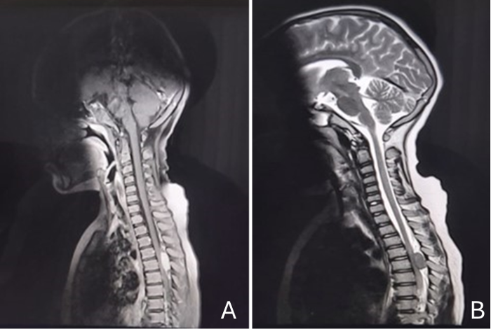
an MRI, CE-MRI, and PET scan, revealing a
well-defined solitary extradural lesion between
DV4-DV6 (measuring 11x25x24 mm), with cortical
erosion noted in the right pedicle of DV5-DV6.
(Fig- 1)
|
| Figure
1 (A and B): Well-defined solitary solid
extra-dural lesion seen at level of lower
border of DV4 to upper border of DV6
vertebrae, epicentered in posterior
epidural space. |
|
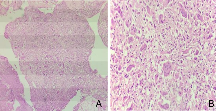
| Figure 2 (A and B):
Scattered multinucleated giant cells
within a mononuclear stroma, consistent
with GCT. |
Histopathological
examination showed scattered multinucleated giant
cells within a mononuclear stroma, consistent with
GCT. (Fig-2) Following laminectomy, the patient
showed clinical improvement, with lower limb
strength returning to a power of 4-5.
Discussion
Giant cell tumors
(GCT) account for 5% of primary bone tumors, with
1-1.5% occurring in the mobile spinal segments.
Among spinal GCTs, the thoracic region is the most
frequently affected, though such occurrences are
rare in children.[1,2]
Histologically, GCTs
feature multinucleated giant cells within a stroma
of round to spindle-shaped mononuclear cells. In
this case, biopsy and histological analysis
confirmed the diagnosis of GCT, ruling out other
possibilities such as aneurysmal bone cysts.[3-6]
Surgical excision
remains the treatment of choice for spinal GCTs,
though it is often challenging due to proximity to
the spinal cord and risk of neurological deficits.
In this case, total tumor excision was achieved
without recurrence noted during follow-up.[3,7-9]
Conclusion
Giant cell tumors of
the thoracic vertebra are uncommon in pediatric
patients, and this case illustrates the importance
of early surgical intervention. The patient
experienced full neurological recovery after the
excision, with no recurrence observed at the
one-year follow-up.
References
- Alfawareh MD, Shah ID, Orief TI, Halawani MM,
Attia WI, Almusrea KN. Pediatric Upper Cervical
Spine Giant Cell Tumor: Case Report. Global
Spine J. 2015 Aug;5(4):e28-33. doi:
10.1055/s-0034-1396433.
- Zabalo G, Ortega R, Vázquez A, Carballares I,
Díaz J, Portillo E. Tumor de células gigantes de
raquis lumbar. Caso clínico y revisión de la
literatura [Giant cell tumor of the lumbar
spine. Case report and review of the
literature]. Neurocirugia (Astur). 2015
Sep-Oct;26(5):251-5. Spanish. doi:
10.1016/j.neucir.2014.11.008.
- Werner M. Giant cell tumour of bone:
morphological, biological and histogenetical
aspects. Int Orthop 2006;30(6):484–489
- Dahlin DC. Giant-cell tumor of vertebrae above
the sacrum: a review of 31 cases. Cancer
1977;39(3):1350–1356
- Çomunoğlu N, Kepil N, Dervişoğlu S.
Histopathology of giant cell tumors of the bone:
With special emphasis on fibrohistiocytic and
aneurysmal bone cyst like components. Acta
Orthop Traumatol Turc. 2019
Jan;53(1):35-39. doi:
10.1016/j.aott.2018.10.007.
- Verschoor AJ, Bovée JVMG, Mastboom MJL, Sander
Dijkstra PD, Van De Sande MAJ, Gelderblom H.
Incidence and demographics of giant cell tumor
of bone in The Netherlands: First nationwide
Pathology Registry Study. Acta Orthop.
2018 Oct;89(5):570-574. doi:
10.1080/17453674.2018.1490987.
- Yuan B, Zhang L, Yang S, Ouyang H, Han S,
Jiang L, et al. Imaging Features of Aggressive
Giant Cell Tumors of the Mobile Spine:
Retrospective Analysis of 101 Patients From
Single Center. Global Spine J. 2022
Sep;12(7):1449-1461. doi:
10.1177/2192568220982280.
- Strøm TM, Skeie AT, Lobmaier IK, Zaikova O.
Giant Cell Tumor: A Rare Condition in the
Immature Skeleton-A Retrospective Study of
Symptoms, Treatment, and Outcome in 16 Children.
Sarcoma. 2016; 2016:3079835. doi:
10.1155/2016/3079835.
- Metkar U, Wardak Z, Katz DA, Lavelle WF. Giant
cell tumor of a lumbar vertebra in a 7-year-old
child: a case report. J Pediatr Orthop. 2012
Dec;32(8):e76-80. doi:
10.1097/BPO.0b013e31826193e7.
|


